
Topic outline
-
There are three main sections in the Darkfield Microscopy course. It starts with creating a system that meets the needs of the practitioner. Those who have some experience can skip most of the Getting Started lesson. So, the broader categories of what is covered are:
- Configuration of a system that fits the budget and clinical or research requirements of the practice, assistance setting up the system for long-distance collaboration, discussion of accessories and sampling techniques, and private tutoring that addresses the nuances of centering, focusing, and skillful use of the microscope, camera, monitor, and record-keeping system for documenting case histories.
- Practice involving identification and correction of weaknesses and problems found; plus discussion of treatment options and interactions between diet, herbs, supplements, and/or prescription medications. Where indicated, toxins, lifestyles, and environmental issues such as exposure to electromagnetic interferences with physiological functioning are also addressed.
- Mastering the use of natural remedies to perfect the plasma, erythrocytes, and white blood cells.
The course includes charts and written material as well as one-on-one sessions with Dr. Ingrid Naiman. There is a forum for discussion that is regulated by the level of enrollment. There is also a secure area for uploading images and case histories that has a gallery for posting that has controlled access.
This course has prerequisites and requires a signed intellectual property agreement.
In some cases, additional charges will apply if the practitioner requests evaluations that go beyond what might be termed classroom as contrasted with clinical consultations.
Enrollment began in June 2022, and applications are currently being accepted.
-
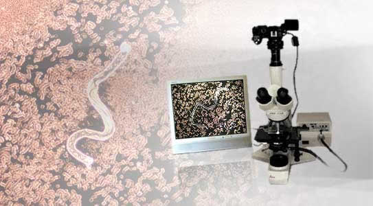
 The first step in getting started with darkfield microscopy is to make sure that the system one is acquiring is configured to one's needs. Once deciding on the model and camera, one needs a few basic supplies and usually some assistance in setting up the system and learning to use it. Mistakes can be costly so it pays to have some help with the assembly and proper use of the equipment.
Next, of course, comes the sampling and there are some tips for preparing the slides and coverslips as well as how to take the best sample possible. Then, the sample is placed on the microscope stage and explored. Depending on one's background, what one does and does not actually observe can differ. Being a medical doctor, hemotologist, pathologist, or microscopist does not necessarily prepare one for interpreting what is seen in darkfield.
The first step is to acquire a microscope so the Getting Started pdf will be sent when the student enrolls. This will be followed by private tutorials. The first real lesson will begin once the scope is in use.-
FileDarkfield Studies Getting Started - by Dr. Ingrid Naiman FileNot available unless: You are a(n) Student
-
-
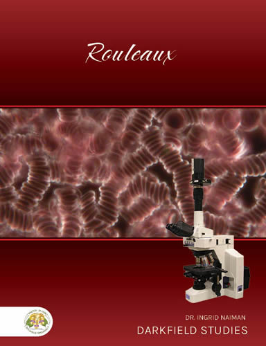 Rouleaux is often the first feature that students of darkfield microscopy identify. The word, rouleaux, comes from the French term for a stack of coins and is actually the technical term used by microscopists.
There are numerous different causes for rouleaux as well as varying symptoms and risks. In this lesson, the factors contributing to rouleaux as well as the corrections are covered in detail. The course includes downloadable material, case histories, dietary suggestions, and herbal remedies for different underlying causes.
Course Date: 7 January 2023, 10 am PST
-
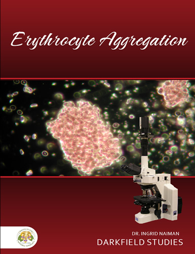 Erythrocyte aggregation is different from rouleaux and may not be as widely distributed, but it is potentially quite dangerous. This involves clustering of cells that are tightly packed. It can lead to thrombosis. There are numerous potential causes ranging from diet, medications, viscosity of the plasma, infection or pathological conditions in the plasma or red blood cells, deformity of cells, pressure on veins and arteries, circulatory issues, and sometimes parasitization. Risks are higher when there is dehydration such as when flying at high altitude and sitting for hours without much movement. It can happen for other reasons such as cells sticking together because of damage to the surface membranes . . . which may, in turn, be due to chemical toxicity, very low zeta potential, or perhaps EMF interference with normal physiological functioning.
The causes along with suggestions for treatment are part of this lesson.
Course Date: to be announced. -
The surface of the erythrocytes has a negative electrical charge so the cells should repel each other. Theories of exactly how this works vary somewhat between disciplines. Hematologists tend to put the emphasis on the specific properties of the sialic acid on the membranes of the red blood cells. Nutritionists often put a bit more emphasis on the hemoglobin inside the cells. There is wide agreement that normal cells will keep their distance from each other and that cells with low zeta potential will benefit from diets that correct for deficiency conditions.
Course Date: To be announced -
There are many types of blood parasites. Some are intraerythrocytic, meaning they live mainly inside red blood cells. However, when they mature, the red blood cells rupture and cause symptoms such as are associated with malaria and babesia. Other parasites live mainly in the plasma and forage on nutrients as well as red blood cells. There are about 4000 known types of parasites that affect humans. These vary in eating habits, behavior, toxicity, and risks to hosts.Course Date: To be announced
-
Image and text coming soon.
Course Date: To be announced
-
Though one of the most common sources of metal toxicity is amalgam dental fillings, mercury is just one of the many toxic metals contributing to health challenges. Aluminum and lead are also very common sources of metal toxicity, but arsenic, cadmium, and barium as well as other metals can cause serious risks, usually heavily impacting the immune system. In darkfield microscopy, the evidence of toxicity is very apparent, but the exact cause has to be determined using other diagnostic methods. Being aware of the underlying causes of toxicity and having the ability to monitor the benefits of chelation therapies is one of the many benefits of studying live blood.Course Date: To be announced
-
Image and text coming soon.
Course Date: To be announced